Maxime Obadia
Radial shock waves
Principle:
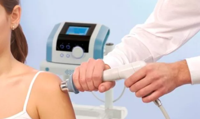
The shockwave device we use, chatTANOOGA/DJO GLOBAL’s Shockwave, A U.S. company specializing in state-of-the-art health devices, produces so-called radial shock waves (RSWT), i.e. outgoing waves will diverge from their source, in “fan”, unlike shock waves, so-called “frontal” waves, which will be linear waves, therefore much more powerful and invasive, but which remain reserved for surgery.
These pneumatic shock waves (air compressor) are delivered on contact with the skin and penetrate the tissues to a depth of 3 to 4 cm, thus crossing the different layers of the skin (skin), fat (fat), muscle or tendon, or even to joint structures, ligaments and capsules, to reach the bone or cartilage. Of course, the intensity of the waves can be adjusted according to the patient’s sensations and the state of inflammation of the structure to be worked.
RSWTs can therefore treat soft tissue lesions mainly
In the early 1990s, machines used in urological environments have expanded their scope to treat pseudarthritis or to fragment intratendinous calcifications etc.

What to treat:
Shock waves can treat all tendonopathies, namely tendonitis, or inflammation of the tendon, tenosynovitis, or inflammation of the tendon sheath, enthesopathies, or inflammation of the bone/tendon junction, or finally tendon/muscle junctions.
They will also be able to treat tendons, bursitis, capsulitis, pseudarthritis, calcifications… some examples
: At the shoulder
- tendonitis of the shoulder and rotator cuff
- calcifying shoulder tendinitis
At the upper limb level
- elbow epicondylite or tennis elbow, elbow epitrochleitis or golf elbow
- Quervain tenosynovitis of the wrist, Dupuytren’s disease (waves will act on tendon retraction)…
At the hip
- middle gluteal tendinitis, pyramidal
- trochanterian bursitis
At the lower limb level
- rotulian tendonitis
- tibial periostitis
- achileca tendinitis and plantar aponévrositis, calcaneal spine
Mode of action:
The chemical action is partly explained by the anaesthetic effect that appears during the session and persists a few hours after the session. After a certain quota of percussion, there would likely be a local release of endorphins or pain-inhibiting substances.
Gate control: The reduction of painful perception is achieved by stimulating large sensitive nerve fibers in the skin, resulting in an inhibition of painful alows in the bone marrow.
Mechanical action: Local inflammation occurs with activation of circulation and tissue repair processes. It has been shown, by doppler echo, that hypervascularization appears after shock wave therapy at the rotator cuff.
With regard to their use in soft tissues, their action is similar to physical therapy techniques (MTP, defibring massage, ultrasound).
Shock waves create micro lesions that can heal better in a second phase (this is the transformation of an area of chronic inflammation into an area of acute inflammation).
The effectiveness of the treatment should be assessed at the end of the last shock wave session and 45 days later.

Protocol:
Treatment is short-term, 4 to 5 sessions maximum, at a rate of 1 to 2 sessions per week; the frequency varies from 0 hz to 20 hz, depending on the pathology: the number of strokes per session varies and can reach 5000. There are 2 tips of 20 mm and 40 mm.
The pressure delivered by the compressor is between 0 and 5 bars.
Success is almost systematic in terms of relieving or decreasing pain. The device allows us to detect the area inflamed by the pain that the waves provide, but which diminishes as we go along…
Healing is achieved in 75% of cases depending on sites and seniority.
The contraindications:
There are few contraindications (pregnancy, vascular or tumor pathologies, local infections, bleeding disorders or anticoagulant treatment).
The side effects :
CDO sessions are sometimes painful but must remain tolerable by the patient.
Delayed side effects are usually of three types: temporary exacerbation of pain, local redness and edema, bruising (usually occurring in areas where the adipose panicle is important).
They are still minors, never prohibit further treatment and are observed in only 10 to 20% of cases.